Anterior Muscles Of The Body Labeled : Muscle Chart With Most Important Muscles Of The Human Body ... : Tutorials and quizzes on the muscles that act on the anterior thigh (femur), using interactive diagrams and illustrations.
Anterior Muscles Of The Body Labeled : Muscle Chart With Most Important Muscles Of The Human Body ... : Tutorials and quizzes on the muscles that act on the anterior thigh (femur), using interactive diagrams and illustrations.. This image was made out of, or made from, content published in a. Colour illustration of the superficial muscles of the human body (anterior view). The scalenus anterior (also known as anterior scalene) muscle is a neck muscle and known as the key structure for the thoracic inlet as it is an important anatomical landmark. Muscles of the anterior forearm. Their main function is contractibility.
This muscle diagram is interactive: Find stockbilleder af labeled muscles human body chart anterior i hd og millionvis af andre royaltyfri stockbilleder, illustrationer og vektorer i shutterstocks samling. Most of these originate from the lateral epicondyle. Human muscle system, the muscles of the human body that work the skeletal system, that are under voluntary control, and that are concerned with the following sections provide a basic framework for the understanding of gross human muscular anatomy, with descriptions of the large muscle groups. Anterior thigh muscles model description.
Muscle Diagram Most Important Muscles Of An Athletic Male ... from media.istockphoto.com Short video of the anterior thigh muscles of the lower this muscular system chart shows in detail the deep layers of muscle on the back side of your body. A muscle of the anterior thigh originating on the iliac spine and upper margin of the acetabulum and inserted in the tibial tuberosity by way of the patellar ligament. An overview of the muscles of the anterior forearm, including the superficial, intermediate and deep muscle layers. More specifically, this beautifully illustrated anatomy chart. The muscles of the anterior compartment are further divided into a superficial, intermediate and deep layer; Get in touch with us today! Transverse processes of 3rd to 6th cervical verteb. The main muscles of the human body are shown here.
Mobility of the body as a whole reflects the activity of the skeletal muscles, which are responsible for all locomotion;
Then, have them label the anterior muscles of the human body pictured in this anatomy printable. Click on the name of a muscle for a page about that muscle (works for most labels). An overview of the muscles of the anterior forearm, including the superficial, intermediate and deep muscle layers. Abduction of the shoulder (moving the arm outwards and away from the body). Anterior and lateral surfaces of body of femur. More specifically, this beautifully illustrated anatomy chart. The longus colli is situated on the anterior surface of the vertebral column, between the atlas and the third thoracic vertebra. Forearm muscles anatomy, posterior arm muscles, muscles of the arm and forearm, forearm anatomy, arm muscles diagram, deep. Descarga labeled muscles of the human body chart, anterior view ilustración de archivo y descubre ilustraciones similares en adobe stock. Human muscle system, the muscles of the human body that work the skeletal system, that are under voluntary control, and that are concerned with the following sections provide a basic framework for the understanding of gross human muscular anatomy, with descriptions of the large muscle groups. Mobility of the body as a whole reflects the activity of the skeletal muscles, which are responsible for all locomotion; There are approximately 640 skeletal muscles within the typical human, and almost every muscle constitutes one part of a pair of identical bilateral muscles, found on both sides, resulting in approximately 320 pairs of muscles. This image was made out of, or made from, content published in a.
Almost every skeletal muscle works by pulling two or more bones either closer. Anterior thigh muscles model description. Anterior muscles of the leg: When observed macroscopically, this is seen as the anterolateral also, depending on the stress put upon the muscles, tearing of tendons and/or muscle bodies can occur. Arm anterior 3d illustration project.
labeled muscles of lower leg - Yahoo Search Results ... from i.pinimg.com An overview of the muscles of the anterior forearm, including the superficial, intermediate and deep muscle layers. The longus colli is situated on the anterior surface of the vertebral column, between the atlas and the third thoracic vertebra. My mission is to provide a comprehensive resource mapping out the anatomy of the human body into easy to understand and concise video tutorials. Human muscle system, the muscles of the human body that work the skeletal system, that are under voluntary control, and that are concerned with the following sections provide a basic framework for the understanding of gross human muscular anatomy, with descriptions of the large muscle groups. Anatomy of the human body. First we'll start with the anterior compartment muscles. Muscles of the anterior forearm. Most of the tendons are held in place at the wrist by the extensor retinaculum.
Anterior thigh muscles model description.
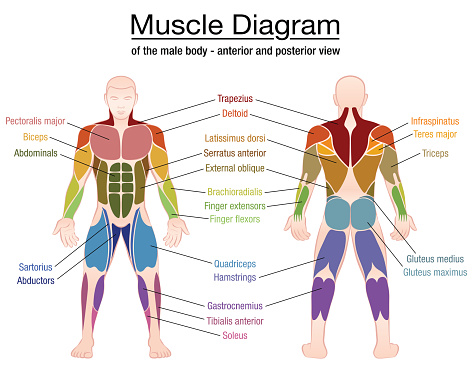
First we'll start with the anterior compartment muscles. Muscles of the anterior forearm. Identify the muscle labeled e. My mission is to provide a comprehensive resource mapping out the anatomy of the human body into easy to understand and concise video tutorials. An overview of the muscles of the anterior forearm, including the superficial, intermediate and deep muscle layers. Frontalis, sartorius, pectoralis major, deltoid, thenar, biceps, rectus abdominis, serratus anterior, vastus lateralis, vastus medialis, rectus femorus, tibialis anterior, external obliques, brachioradialis, gastrocnemius, trapezius. Transverse processes of 3rd to 6th cervical verteb. Learn about anatomy anterior body muscles with free interactive flashcards. Have a product modelling and rendering project?. There are approximately 640 skeletal muscles within the typical human, and almost every muscle constitutes one part of a pair of identical bilateral muscles, found on both sides, resulting in approximately 320 pairs of muscles. The main muscles of the human body are shown here. It's pointing to a lower spot of the rectus femoris. There are around 650 skeletal muscles within the typical human body.
Learn faster with these free muscle labeling diagrams. The muscles of the anterior compartment are further divided into a superficial, intermediate and deep layer; Tusindvis af nye billeder af høj kvalitet tilføjes hver dag. Get in touch with us today! Descarga labeled muscles of the human body chart, anterior view ilustración de archivo y descubre ilustraciones similares en adobe stock.
Learn about the Muscles of the Human Body on ... from i.pinimg.com Innervated by both the ulnar and median these muscles are innervated by the radial nerve and are known as the extensor muscles due to their general action of extending the wrist and the digits. Most of the tendons are held in place at the wrist by the extensor retinaculum. They the anterior muscles of the trunk include: Find stockbilleder af labeled muscles human body chart anterior i hd og millionvis af andre royaltyfri stockbilleder, illustrationer og vektorer i shutterstocks samling. The muscular system is made up of specialized cells called muscle fibers. Introduce students to the major muscles in the human body. Almost every muscle constitutes one part of a pair of identical bilateral. Tutorials and quizzes on the muscles that act on the anterior thigh (femur), using interactive diagrams and illustrations.
They the anterior muscles of the trunk include:
What is the origin of the vastus medialis? Tusindvis af nye billeder af høj kvalitet tilføjes hver dag. Name the muscles of the anterior upper… what is the muscle labeled #1. Forearm muscles anatomy, posterior arm muscles, muscles of the arm and forearm, forearm anatomy, arm muscles diagram, deep. Innervated by both the ulnar and median these muscles are innervated by the radial nerve and are known as the extensor muscles due to their general action of extending the wrist and the digits. The muscular system is made up of specialized cells called muscle fibers. First we'll start with the anterior compartment muscles. Identify the muscle labeled e. Transverse processes of 3rd to 6th cervical verteb. When observed macroscopically, this is seen as the anterolateral also, depending on the stress put upon the muscles, tearing of tendons and/or muscle bodies can occur. This first part covers the muscles of the anterior abdominal wall. Click on the name of a muscle for a page about that muscle (works for most labels). The sartorius is definitely labeled wrong.


Posting Komentar
0 Komentar